Principal Investigators
Mission Statement of the Lab
General Overview and Areas of Focus
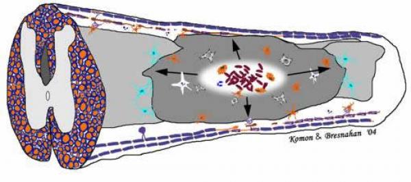
The CNS response to injury shares features with other organ systems and includes surprisingly robust repair mechanisms. The cascade of events that leads to dysfunction and partial recovery after spinal cord injury, for example, includes overlapping degenerative and regenerative processes mediated by inflammation, excitotoxicity, growth and trophic factor production in all of the cellular components of the CNS including neurons, glia, immune cells, and vascular elements.
In this complex cascade, neural connections may be broken and remade and the final functional outcome reflects the resolution of all of these interacting events. We think that a better understanding of the biology of the lesion and repair processes will lead to better strategies for providing protection against secondary injury, enhancing self-repair, and for engineering repair through the use of cellular implants and biomaterials.
We are invested in the production of useful in vitro and in vivo testing systems for bringing these strategies into the clinic. The laboratory has recently moved from The Ohio State University to UCSF (November, 2006)
Accomplishments
Our group is known for developing preclinical models to study the recovery of function after spinal cord injury, and for studies of the biology of neural injury and repair.
Current projects
- Analyses of the interrelationship of post-injury inflammatory events and excitotoxic cell death
- The roles of oligodendrocyte death and replacement in recovery after injury
- The development of stem and progenitor cell transplantation strategies for promoting recovery after spinal cord injury
The laboratory continues to develop new animal paradigms for modeling human neurotrauma. BASIC's location at the SFGH along with the physician-scientists of UCSF Neurotrauma program will promote basic and clinical science interactions, and new animal models will be generated based on knowledge of current treatments and outcomes in the human neurotrauma arena. For example, there is an emerging interest in post injury edema, studied in rodents using high field MR imaging. Acute studies of interventions that can reduce edema in animal studies may be taken directly to clinical studies in the neurotrauma ICU. Our goal is to help translate the laboratory's expertise in the biology of injury and recovery to treatments that can be implemented and tested in neurotrauma patients at SFGH and other centers.
Current Objectives
Transplantation of stem-like and precursor cells into spinal cord contusion lesions: This is currently a major thrust in the laboratory. Cells derived from a transgenic rat with ubiquitous expression of the human placental alkaline phosphatase (hPlAP) gene are isolated form embryonic spinal cord and treated with growth factors to produce a glial restricted precursor population. Transplantation into our well-established models of spinal cord injury allows assessment of survival, phenotype, and possible effects on a variety of neurological outcome measures. These cells survive even when transplanted immediately after an injury, and some form astrocytes and oligodendrocytes. The rodent models in our laboratory are also suitable for proof of concept studies using human ES cells (see future directions). Collaborators include Mark Noble, Margot Mayer-Proschel, and Chris Proschel (University of Rochester). We have recently developed a procedure for producing consistent unilateral cervical contusion injuries in rats, and this lesion will be used in much of our proposed work.
Demyelination and remyelination:
We continue to study secondary degeneration of axons and the effects on oligodendrocytes and myelin. The hypothesis is that long-term apoptotic death of myelinating cells reduces the efficiency of conduction in spared axonal tracts after injury, and that saving these cells and/or producing remyelination through stimulation of endogenous stem cells or implantation of glial precursors will improve recovery from CNS injury. The secondary cell death may be due to a variety of receptor-mediated cascades, e.g. the activation of TNF receptors and p75 by endogenous ligands (e.g. Beattie et al, 2002). Recently, we have discovered that axonal degeneration can induce oligodendrocyte precursor cell proliferation and differentiation. However, in the presence of oxidative stress, new cells are apparently lost via apoptosis. The role of microglial activation in these events is being studied both in vitro and in vivo. These projects overlap with those on inflammation and excitotoxicity, and also have relevance for other neurological disorders, including multiple sclerosis and stroke. Collaborators include the New York State consortium group (see below)
Inflammation and excitotoxicity:
We have found that the pro-inflammatory cytokine, TNF-ehances glutamate-receptor-mediated excitotoxic cell death in neurons in vitro and in vivo. This is due, at least in part, to changes in AMPA receptor trafficking to the cell surface (see E. Beattie et al, 2002). Confocal microscopy techniques have been developed to track the surface expression of these receptors in vivo after injury or nano-injections of substances. Collaborators include Eric Beattie at CPMC.
Future Outlook
Imaging transplanted cells: Transplanted stem and precursor cells can be transfected with EGFP and also labeled with paramagnetic particles (e.g. iron oxide). We have completed studies of spinal cord contusion lesions using a 4.7T small bore MRI and plan to extend these observations to transplanted cells in the near future. This project is intended to provide the beginnings of techniques for longitudinal tracking of cellular implants to detect viability and migration, using novel contrast agents and imaging techniques. Collaborators included Drs. Petra Schmalbrock and Michael Knopp (Radiology at OSU).
The role of AMPA receptors in oligodendrocyte differentiation and response to injury: Our studies of AMPA receptor trafficking after neuronal injury will be complimented by work on oligodendrocyte precursor cells, and mature oligodendrocytes in vitro and in vivo after injury. The hypothesis is that cytokines and glutamate interact via cytokine-mediated alterations in AMPA receptor sensitivity (e.g. increased surface expression).
Preconditioning stem cells for implantation: We have received a supplemental award to an NIH grant that will fund the development of preconditioning strategies for human ES cells. We are exploring the cell lines available for use. The strategy will be to pre-differentiate cells to produce glial-restricted populations, and to treat with antioxidant therapies to enhance survival after implantation. Collaborative tests of cellular and pharmacologic therapieshave recently been funded, with our laboratory providing lesion models and outcomes measures for therapies involving cAMP plus cellular implants (New York State Spinal Cord Research Program consortium, and Center for Neural Repair at UCSD). Our laboratory continues to work on developing useful models of CNS injury for translation to preclinical, and eventually clinical, studies of cellular and pharmacologic therapies. We are also involved in the development of robotics systems for enhancing recovery of function in rat models of SCI, with Neville Hogan and colleagues at MIT.
Funding and Contributors
Funding for the Laboratory and its member s has come from:
NIH (NINDS)
The New York State Department of Health and Burke Rehabilitation Institute
The C.H. Neilsen Foundation
The Roman Reed Program
International Spinal Research Trust
Christopher Reeve Foundation
The Paralysis Project of America
Paralyzed Veterans of America
Current Lab Members
J Russell Huie, PhD (Postdoctoral Scholar)
Amity Lin, BS (Staff Research Associate II)
Xiaokui Ma, MD (Core Histopathology Specialist)
Kate Mitchell, (Graduate Student Volunteer)
Payam Saadai, MD (Surgical Resident)
Jason Talbott, MD, PhD (Radiology Resident)
Jinghua Yao (Core Histopathology Staff Resea rch Associate)
Recent laboratory members at OSU and UCSF:
Caitlin Hill, Ph.D. (now at University of Miami)
Randy Christensen, Ph.D. (now at Coe College)
John Gensel, Ph.D. (now at Ohio State University)
Fang Sun, Ph.D. (now at Harvard/ Children's Hospital)
Gregory Holmes, Ph.D. (now at Pennington Research Institute)
Byeong-Keung Ha, M.D., Ph.D (now at Case Western Reserve University SOM)
Sergio Veiga, PhD (now at Universidad Alfonso X el sabio Madrid, Spain)
Karen-Amanda Irvine, PhD (now at Stanford University)
Brandon A Miller, MD/PhD (now at Emory University)
Collaborators
The Ohio State University: Georgeta Mihai, Ph.D., Petra Schmalbrook, Ph.D., Dept. of Radiology.
University of Rochester: Mark Noble, Ph.D.; Margot Mayer-Proschel, Ph.D.; Chris Proschel, Ph.D.
UCSD, UCLA, UCI, UCD, Mark Tuszyn ski, M.D., Ph.D. et al, California Primate Consortium.
California Pacific Medical Center Research Insititute: Eric Beattie, Ph.D.New York State Spinal Research Program Consortium - Raj Rattan, M.D., Ph.D. (Cornell/Burke Rehabilitation Center); Mark Noble, Ph.D. (Rochester); Barbara Hempstead, Ph.D. (Weill/Cornell); Marie Filbin, Ph.D. (Hunter College); Neville Hogan (MIT) and teams at Rutgers University and Acorda Therapeutics.
Selected Publications
Crowe, M.J., Bresnahan, J.C., Shuman, S.L., Masters, J.N., and Beattie, M.S. (1997) Apoptosis and delayed degeneration after spinal cord injury in rats and monkeys. Nature Med., 3: 73-76.
Beattie MS, Rogers RC, Hermann GH, Bresnahan JC (2002) Cell death in models of spinal cord injury, Prog. Brain Res. 137: Chapter 4 , pp 37-47 (Volume title, Spinal Cord Trauma: Regeneration, Neural Repair and Functional Recovery‚ edited by L. Mckerracher, G. Doucet and S. Rossignol).
Beattie EC, Stellwagen D, Morishita W, Bresnahan J, Ha B-K, Von Zastrow M, Beattie MS*, Malenka RC* (2002) Control of synaptic strength by glial TNFalpha. Science, 295: 2282-2285. *co-corresponding authors.
Beattie MS*, Harrington AW*, Kim MS, Boyce SL, Longo F, Hempstead BL, Bresnahan JC, Yoon SO (2002) Pro-NGF induces p75-mediated death of oligodendrocytes following spinal cord injury, Neuron, 36: 375-386. *co-1st authors
Hill CE, Proschel C, Noble M, Mayer-Proschel M, Gensel JC, Beattie MS, Bresnahan JC (2004) Acute Transplantation of glial restricted precursor cells into spinal cord contusion injuries: Survival, differentiation and effects on lesion environment and axonal regeneration. Exp. Neurol., 190: 289-310.
Beattie MS (2004) Inflammation and apoptosis: linked therapeutic targets for spinal cord injury. Trends in Molecular Medicine, 10: 580-583.
Miller BA, Sun F, Christensen RN, Ferguson AR, Beattie MS, Bresnahan JC (2005) A sublethal dose of TNF potentiates kainite-induced excitotoxicity in optic nerve oligodendrocytes. J. Neurochem. Res., 30: 867-875.
Gensel JC, Tovar CA, Hamers FPT, Deibert RJ, Beattie MS, Bresnahan JC (2006) Behavioral and histological characterization of unilateral cervical spinal cord contusion injury in rats. J Neurotrauma, 23: 36-54.
Christensen RN, Ha BK, Sun F, Bresnahan JC and Beattie MS (2006) Kainate induces rapid redistribution of the actin cytoskeleton in microglia. J. Neurosci. Res., 84: 170-181.
Miller BA, Crum JM, Tovar CA, Ferguson AR, Bresnahan JC, Beattie MS (2007) Activated microglia reduce olidodendrocyte progenitor cell viability but protect mature oligodendrocytes from apoptotic cell death. J. Neuroinflammation, 4:28 (26 November, 2007) title="ht<mce:script type=">tp://www.jneuroinflammation.com/" href="http://www.jneuroinflammation.com/">http://www.jneuroinflammation.com/
Nout YS, Bresnahan JC, Culp E, Tovar AC, Beattie MS, Schmidt MH (2007) Novel technique for monitoring micturition and sexual function in male rats using telemetry. Am J Physiol Regulatory Integrative Comp Physiol, 292: R1359 - R1367.
Mihai G, Nout YS, Tovar CA, Miller BA, Schmalbrock P, Bresnahan JC, Beattie MS (2008) Characterization and comparison of two severities of cervical spinal cord injury in rats using magnetic resonance imaging. J Neurotrauma, 25: 1-18.


